Hip Groin Muscle Anatomy / Common Groin Injuries My Family Physio / In human anatomy, the muscles of the hip joint are those muscles that cause movement in the hip.
Hip Groin Muscle Anatomy / Common Groin Injuries My Family Physio / In human anatomy, the muscles of the hip joint are those muscles that cause movement in the hip.. The location of the center of the entire axis is at the femoral head. The femur, the hip bone (subdivided into ilium, ischium and pubis) as well as the sacrum were labeled separately with differently colored labels The different anatomical areas of the gluteal region: This group includes the adductor magnus, adductor longus, and adductor brevis muscles, as well as the pectineus and gracilis. Über 7 millionen englischsprachige bücher.
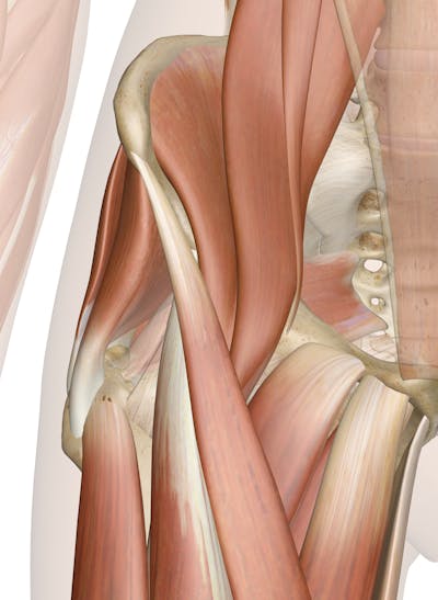
The hip bone, also known as the innominate bone, coxal the iliopsoas muscle is really a combination of 3 muscles: Anatomy of the groin area superficial muscles and deep muscles in this image, you will find rectus abdominis, external oblique, inguinal ligament, tensor fascia lata, gracilis, sartorius, rectus femoris, the iliotibial band in it. This group includes the adductor magnus, adductor longus, and adductor brevis muscles, as well as the pectineus and gracilis. There are many muscles located in this region that support and move the hip and pelvis. The gluteals are the muscles in your buttocks.
Tendinitis And Bursitis Treatment Cincinnati Tendinitis Dayton Oh from www.beaconortho.com This is also known as the medial compartment of the thigh that consists of the adductor muscles of the hip or the groin muscles. Groin strain, also referred to as a pulled groin muscle, typically occurs as a result of an athletic injury or awkward movement of the hip joint, which leads to stretching or tearing of the inner thigh muscles. Anatomy of the groin area superficial muscles and deep muscles in this image, you will find rectus abdominis, external oblique, inguinal ligament, tensor fascia lata, gracilis, sartorius, rectus femoris, the iliotibial band in it. This condition occurs when a bursa becomes inflamed. Pain experienced in the hip, groin and pelvic region may be related to: The adductor muscle group, also known as the groin muscles, is a group located on the medial side of the thigh. Über 7 millionen englischsprachige bücher. They are important for stabilising the body and for moving the legs.
The adductor longus and gracilis both originate form from the pubic bone.
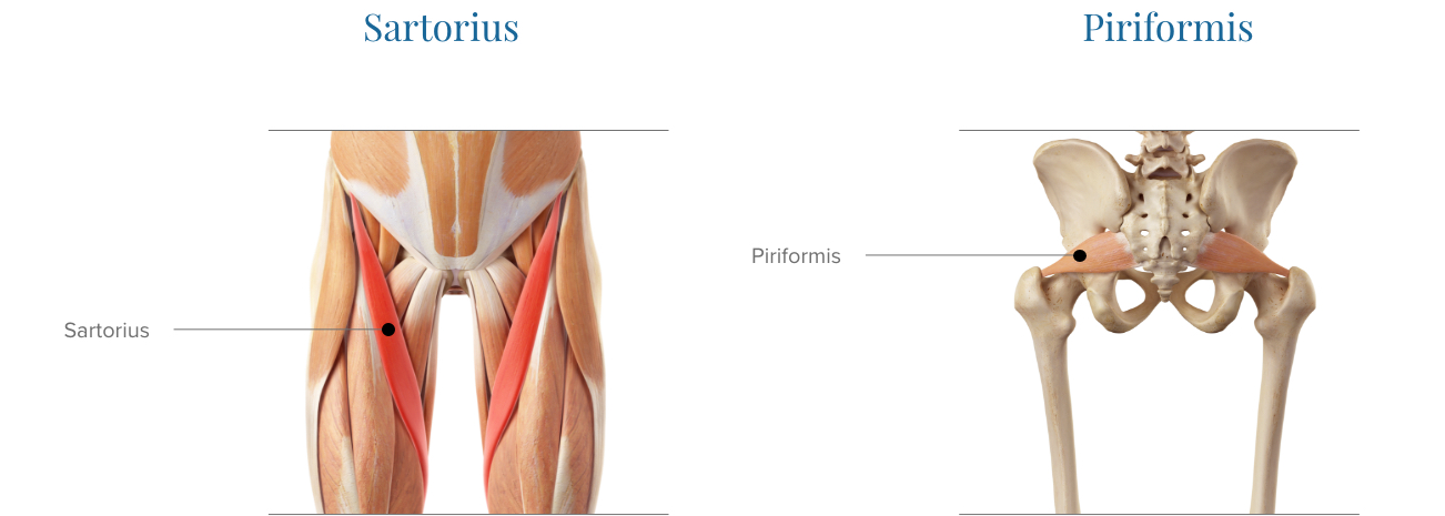
The muscles and the bones are under the layer of subcutaneous fat. Groin muscles diagram anatomy of groin area photos muscles. The gracilis muscle is one of the muscles found in the groin. The largest bursa of the human body, which is called the iliopsoas bursa, is located between the iliopsoas tendon and the pelvis. This group of muscles includes: Blood vessels and nerves of the hip In human anatomy, the muscles of the hip joint are those muscles that cause movement in the hip.most modern anatomists define 17 of these muscles, although some additional muscles may sometimes be considered. The canal is like a tube with 4 sides: The adductor longus and gracilis both originate form from the pubic bone. The hip muscles include pelvic and groin muscles. These muscles move the thigh toward the body's midline. The gluteus medius, gluteus minimus, piriformis, tensor fasciae latae on the outside. The iliopsoas muscle and tendon are responsible for hip flexion (bending or moving forward) and external (lateral) rotation of the hip joint.
Bursitis is one of the most common cause of pain in the hip and groin. This group includes the adductor magnus, adductor longus, and adductor brevis muscles, as well as the pectineus and gracilis. The groin muscles are a group of muscles situated high on the leg in the inner thigh. Gluteus medius in pink and minimus in blue. Healthy bursae reduce friction between bones and tendons or between muscles, but repetitive motions can irritate bursae and contribute to bursitis.
Muscles Of The Hip Anatomy Pictures And Information from innerbody.imgix.net In human anatomy, the muscles of the hip joint are those muscles that cause movement in the hip. The groin is the area that lies between the abdomen (stomach) and thighs. The gluteus medius, gluteus minimus, piriformis, tensor fasciae latae on the outside. Your hip joint is found along the same line underneath your groin. Here we explain the hip and groin muscles, their actions and exercises. Front layer (external oblique fascia), back layer (posterior wall), medial layer (straight abdominal muscles) and lateral layer (inguinal ligament). Groin muscles diagram anatomy of groin area photos muscles of the groin diagram human. Lateral rotation is needed for crossing the legs.
Gluteus medius in pink and minimus in blue.
Anatomy of the groin area superficial muscles and deep muscles in this image, you will find rectus abdominis, external oblique, inguinal ligament, tensor fascia lata, gracilis, sartorius, rectus femoris, the iliotibial band in it. The largest bursa of the human body, which is called the iliopsoas bursa, is located between the iliopsoas tendon and the pelvis. These muscles move the thigh toward the body's midline. The iliopsoas muscle and tendon are responsible for hip flexion (bending or moving forward) and external (lateral) rotation of the hip joint. The adductor longus and gracilis both originate form from the pubic bone. Groin muscles diagram anatomy of groin area photos muscles of the groin diagram human. Groin muscles diagram groin muscle anatomy diagram hip upper leg muscles diagram wiring. Lateral rotation is needed for crossing the legs. This is also known as the medial compartment of the thigh that consists of the adductor muscles of the hip or the groin muscles. Pain experienced in the hip, groin and pelvic region may be related to: Effective evaluation of the hip and groin depends upon an understanding of local anatomy and function and the proper performance of a focused physical examination. Hip and groin ultrasound education showing how to, scanning protocol, normal anatomy, anatomic variants, labrum, trochanter, bursa, iliopsoas tendon and hernias. They run from the inner thighs and run up to the pelvic bone.
The gracilis is a superficial muscle of your groin and inner thigh that serves to adduct your hip. Über 7 millionen englischsprachige bücher. Healthy bursae reduce friction between bones and tendons or between muscles, but repetitive motions can irritate bursae and contribute to bursitis. Front layer (external oblique fascia), back layer (posterior wall), medial layer (straight abdominal muscles) and lateral layer (inguinal ligament). Groin muscles diagram anatomy of groin area photos muscles.
Amazon Com Hip Brace For Sciatica Pain Relief Groin Thigh Hamstring Compression Sleeve Support Men Women Si Joint Hip Flexor Labral Tear Arthritis Bursitis Belt Sciatic Nerve Wrap Spica Stabilizer Pull Strain from images-na.ssl-images-amazon.com These muscles move the thigh toward the body's midline. In human anatomy, the groin (the adjective is inguinal, as in inguinal canal) is the junctional area (also known as the inguinal region) between the abdomen and the thigh on either side of the pubic bone. Hip and groin pain are common complaints among active adults and pose a diagnostic challenge to clinicians due to the complex anatomy and biomechanics of the region. This is also known as the medial compartment of the thigh that consists of the adductor muscles of the hip or the groin muscles. This group includes the adductor magnus, adductor longus, and adductor brevis muscles, as well as the pectineus and gracilis. Bursitis is one of the most common cause of pain in the hip and groin. Causes of pain in the hip and groin can be musculoskeletal or internal. The hip muscles include pelvic and groin muscles.
The different anatomical areas of the gluteal region:
The femur, the hip bone (subdivided into ilium, ischium and pubis) as well as the sacrum were labeled separately with differently colored labels Musculoskeletal issues begin in the bones,. Hip and groin pain are common complaints among active adults and pose a diagnostic challenge to clinicians due to the complex anatomy and biomechanics of the region. Groin muscles help support the hip joint. Hip flexion is maximal with a high, forward kick that brings the leg above the level of the waist. Groin muscles diagram anatomy of groin area photos muscles. This group of muscles includes: The different anatomical areas of the gluteal region: The transverse axis permits flexion and extension movement. Healthy bursae reduce friction between bones and tendons or between muscles, but repetitive motions can irritate bursae and contribute to bursitis. They are important for stabilising the body and for moving the legs. The groin canal (inguinal canal) connects the inside with the outside of the abdomen and is an opening in the stomach muscles that contains the spermatic cord. Often groin strain occurs in the area of inguinal ligament.
Hip flexion is maximal with a high, forward kick that brings the leg above the level of the waist groin muscle anatomy. In human anatomy, the muscles of the hip joint are those muscles that cause movement in the hip.most modern anatomists define 17 of these muscles, although some additional muscles may sometimes be considered.


0 Komentar